Keyhole IPOM repair for ventral, incisional and umbilical hernias — the mesh goes inside, completely avoiding your existing scar. Particularly effective for obese patients and complex multi-defect repairs. Performed at Sterling Hospitals, Vadodara with 25+ years of hernia expertise.

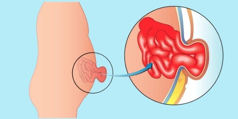
A ventral hernia is a condition where tissue or part of the intestine pushes through a weak spot in the abdominal wall, creating a visible bulge. Many patients experience discomfort, pain, or cosmetic concerns — especially after previous abdominal surgeries, where scar tissue is inherently weaker than normal abdominal wall. While some hernias may appear small and harmless at first, they can gradually enlarge and lead to serious complications if left untreated.
Traditionally, ventral hernias were repaired with open surgery involving large incisions, longer hospital stays, and extended recovery times. Today, laparoscopic ventral hernia repair using the IPOM (Intraperitoneal Onlay Mesh) technique is the preferred approach for most patients. The mesh is placed inside the abdominal cavity through keyhole incisions — completely avoiding any dissection through the existing scar. This produces significantly less pain, faster recovery, and a dramatically lower wound complication rate compared to open repair.
At our practice, we combine over 25 years of surgical expertise, 8,000+ successful laparoscopic surgeries, and advanced technology to deliver world-class hernia repair with a patient-first approach.
A ventral hernia refers to any hernia through the front abdominal wall. Common types include:
Develops at the site of a previous surgical scar — the most common type, occurring in approximately 10–15% of all abdominal operations. Typically appears 1–3 years after surgery.
Occurs at or near the navel (belly button). Common in adults with obesity, multiple pregnancies, or conditions that cause persistent abdominal straining.
Appears between the belly button and the lower chest. Often small but can cause persistent discomfort, especially on bending forward or coughing.
Occurs along the side of the abdominal wall at the semilunar line. Less common but sometimes missed because the bulge is not always visible externally.
All types occur when abdominal muscles weaken, allowing internal tissues to protrude. All require surgical repair — hernias do not resolve on their own.
Ventral hernias do not heal without surgery. Understanding the warning signs and when to seek care — including emergency signs — is critical.
Ventral hernias do not heal on their own. Without treatment, they often enlarge and can lead to incarceration (hernia contents trapped and irreducible), strangulation (blood supply cut off — a life-threatening emergency requiring immediate surgery), or bowel obstruction. Early elective repair is significantly safer than emergency surgery. Do not wait if your hernia is enlarging or causing pain.
Every page on this site is written and medically reviewed by Dr Samir Contractor — a practising fellowship-trained surgeon — not by a content agency. Here is the evidence behind that claim.
Over 25 years of continuous surgical practice at Sterling Hospitals, Vadodara. Every claim on this page is drawn from direct clinical experience — not textbook paraphrasing.
Fellowship-trained at the Royal College of Surgeons of Edinburgh with subspecialty MIS training at Sir Ganga Ram Hospital, New Delhi. Board-certified in multiple countries.
Affiliated with Sterling Hospitals — a leading multi-specialty hospital in Vadodara. Active member of recognised surgical bodies in India, the United Kingdom and the United States.
Transparent pricing published on every procedure page. Surgery recommended only when clinically indicated. 4.9★ patient rating from named, verified patient reviews.
Most patients with ventral or incisional hernias are suitable for laparoscopic IPOM repair. Suitability is confirmed after clinical examination and CT scan of the abdomen to assess defect size and number.
The IPOM technique offers distinct advantages over conventional open repair — especially for incisional hernias, where avoiding dissection through the old scar is the single most impactful difference.
The single biggest advantage of IPOM. No cutting through existing abdominal wall scar dramatically reduces wound infection, seroma, and poor healing — the primary problems with open incisional hernia repair.
Three to four keyhole incisions of 5–10 mm vs a large open wound in scarred tissue. Patients consistently report much less pain than with open repair and are surprised at how comfortable recovery is.
Poor wound healing in large abdominal wounds is a major risk in obese patients after open repair. IPOM avoids this entirely — making it the strongly preferred approach for overweight patients who are at high risk of wound complications.
Discharged within 24–48 hours. Return to desk work in 1–2 weeks. Full activity at 6–8 weeks. Compare to open repair: 6–8 day hospital stay, 6–8 weeks for desk work, 3–4 months for full activity.
Multiple small midline defects (Swiss cheese pattern) can be covered with a single large mesh through the same keyhole approach — something that would require a very long open wound to address surgically.
Recurrence under 5% at 5 years with proper mesh overlap — comparable to or better than open mesh repair — with a significantly lower wound complication rate. Lower infection risk: smaller wounds, less exposure.
Laparoscopic ventral hernia repair uses a laparoscope and specialised instruments through 3–4 small incisions. The mesh is placed inside the abdominal cavity to reinforce the defect from within — the IPOM technique. Here is exactly what happens at every stage.
A detailed assessment ensures the repair is planned precisely before surgery.
Surgery is performed under general anaesthesia — you are completely asleep and comfortable throughout. A urinary catheter is placed for larger repairs. Procedure duration is typically 1–2 hours depending on defect size and the extent of adhesions requiring division.
Three to four ports are placed in the flanks, positioned away from the hernia defect and away from the existing abdominal scar. This is a key principle of the IPOM technique — working from angles that preserve the scar tissue, not through it. A HD laparoscope provides a clear internal view of the entire abdominal cavity.
Any adhesions — scar tissue between the bowel and the abdominal wall near the defect — are carefully divided using cold scissors and energy devices under direct vision. The bowel is fully inspected. The hernia sac and any intestine or omentum within it is reduced back into the abdominal cavity.
A dual-mesh (with an anti-adhesion coating on the intraperitoneal surface to prevent bowel adherence) is introduced through a port and unrolled to cover the defect with a minimum 3–5 cm overlap on all sides. The mesh is secured with transfascial sutures passed through the abdominal wall at the periphery, and additional tackers around the mesh edge. This ensures flat, firm fixation without any folding. Instruments removed, CO₂ released, and port sites closed with absorbable sutures. No drain required in most cases.
Recovery from laparoscopic IPOM is significantly faster than open ventral hernia surgery. Without a large wound to heal, the main limitation is simply allowing the mesh to integrate into the abdominal wall — typically 6–8 weeks.
Oral fluids within a few hours of surgery. Walking within 6–8 hours is actively encouraged and important for preventing post-operative complications including DVT. Mild abdominal bloating from CO₂ gas resolves overnight. Mild pain is well-controlled with oral medication.
Discharged after 24–48 hours. Normal soft diet resumed the morning after surgery — dal, khichdi, curd, soft roti, cooked vegetables. An abdominal binder is provided and should be worn during the day for 4–6 weeks (can be removed for sleeping). Mild pain managed with paracetamol and NSAIDs.
Walking 3–4 times daily, increasing distance each day. Avoid constipation — use stool softeners if needed, as straining increases intra-abdominal pressure and risks mesh displacement in the first weeks. Avoid lifting more than 2–3 kg. Wound dressing review if required.
Most office workers return to light desk duties within 1–2 weeks. Driving is safe when you can perform an emergency stop without discomfort. Full normal Indian diet resumed — dal, roti, sabzi, curd, rice. Continue wearing abdominal binder during the day.
Light gym work, yoga, walking and cycling are all safe. Continue abdominal binder for support during physical activity. Avoid heavy resistance exercises and lifting over 5 kg. Review with Dr Samir to confirm progress.
Gym, sports, heavy manual work and all activities fully resumed. Mesh is well integrated. Final follow-up with Dr Samir. Maintaining a healthy weight and avoiding constipation long-term helps prevent recurrence.
Laparoscopic ventral hernia surgery is very safe. Complication rates are significantly lower than open repair — particularly for wound-related complications which are the major concern with open incisional hernia surgery.
Wound infection at the small port sites is under 1% — significantly lower than the 5–10% wound infection rate after open incisional hernia repair, particularly in obese patients.
A fluid collection under the skin at the old hernia site occurs in 10–15% of cases and usually resolves within 6–8 weeks. Wearing an abdominal binder reduces formation. Large seromas can be aspirated in clinic if symptomatic.
Under 5% at 5 years with proper mesh overlap and fixation — comparable to open repair. Maintained by healthy weight, avoiding constipation, and adhering to lifting restrictions during recovery.
A rare complication (<0.5%) during adhesiolysis. The HD laparoscope provides excellent visualisation, and bowel is fully inspected before port closure. Risk is higher in patients with extensive previous surgery.
Very rare (<0.5%) with modern dual-mesh. Potential issues include mesh infection and mesh-bowel erosion — both extremely uncommon when an appropriate anti-adhesion coated mesh is used by experienced surgeons.
Required in fewer than 3% of cases — typically when adhesions are too dense to divide safely laparoscopically. Conversion is a surgical decision made for patient safety, not a complication.
With 25+ years of surgical expertise and 8,000+ successful laparoscopic procedures, our team minimises these risks through meticulous CT-guided pre-operative planning, precise technique, and close post-operative monitoring. Complication rates at Sterling Hospitals, Vadodara are consistently below published national averages.
All-inclusive package costs — no hidden charges. Two room categories available. Final confirmation after CT scan review and consultation.
| Laparoscopic Ventral / Incisional Hernia Repair (IPOM) | ₹1,00,400 | ₹1,55,600 |
| Laparoscopic Umbilical Hernia Repair | ₹1,00,400 | ₹1,55,600 |
Very large defects or complex multi-sinus repairs may incur additional costs — confirmed at consultation after CT scan review.
WhatsApp gets the fastest response. For appointment booking by phone, call the Sterling Hospital reception during OPD hours.
Popular searches